
1
Negotiate Terms with Seller
Discuss and agree upon the equipment's condition, configuration, and pricing with the seller before proceeding.
| Brand | TOPCON |
| Condition | Refurbished |
| Model | 3D OCT-2000 |
| Warranty | Not specified |
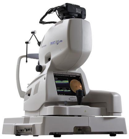
Used Topcon 3D OCT-2000 w/Digital Non-Myd Retinal Camera
The Topcon 3D OCT-2000 System is the first
Spectral Domain OCT system to incorporate a high resolution fundus camera and a userfriendly
color touch screen display in a compact, space saving design.
What makes the Topcon 3D OCT-2000 unique:
» Integrated, high resolution (12.3MP) fundus camera
» FastMap™ software enables dynamic viewing of 2D, 3D and fundus images simultaneously
» Embedded touch-screen for quick and easy navigation
» Historic patient data from Stratus® OCT can be easily imported, analyzed and viewed
» Seamless integration with EyeRoute® Image Management System
3D OCT Image
Topcon’s proprietary FastMap software pioneered the 3D visualization of OCT data, providing another
dimension of clinical information, thereby enhancing the understanding and illustration of complex
pathologies such as vitreous traction, macular edema and retina schesis.
» 3D visualization of OCT data
» Illustrates complex pathologies
» Quickly export images and 3D movies for
presentations
Fundus Image
High resolution, non-mydriatic, color fundus images allow for visualization of conditions which
otherwise would go undetected with OCT technology, such as disc hemorrhages. The 45 degree
field of view and the availability of stereo photos provides the ultimate diagnostic insight.
» High resolution 12.3 MP color fundus camera
» Easily capture and view stereo photos
» PinPoint Registration™ of OCT data in the fundus image
B-scan Image
FastMap software encompasses the latest in noise reduction algorithms and overlapping scanning
technology to create exquisite B-Scan images, which are available almost instantaneously. This
greatly reduces chair time for the patient and enhances offi ce workfl ow.
» Uses latest in noise reduction algorithms
» Enhances office workflow
» Customize capture protocols
Comprehensive Capturing Capture images of the fovea and optic nerve head in one single
scan and high resolution images of the choroid with automatic choroid reference mode.
Video Functions
Use single touch control to review and playback images
and create 2D and 3D videos.
Extended Scanning Depth
Capture high-quality images of high-myopic and hyperopic
patients with a diopter compensation lens and an extended
scanning depth of 2.3 mm.
Compare Function
Allows you to visualize serial exams or view both eyes side by side.
FastMap Software
Topcon’s proprietary FastMap software ensures consistent, high quality images. Using enhanced 3D registration
technology, FastMap reduces artifacts which may be caused by eye movement. This in turn allows for speedy
PinPoint Registration of the OCT data within the fundus image without sacrifi cing workfl ow and preserves image
quality of the vitreo-retinal interface.
In addition, FastMap provides dynamic, simultaneous viewing of the fundus image, and 2D and 3D OCT data,
and automatically detects ILM, RFNL, IS/OS junction, RPE and Bruch’s membrane, which can also be modified.
Thickness Measurement Functions
FastMap software incorporates the latest layer detection algorithms, allowing you to automatically measure total
retinal thickness, RNFL or compare against your legacy Stratus measurements. Manual adjustment of all measurement
grids combined with auto and manual registration of serial exams gives you the highest level of confi dence in
retinal and RFNL thickness measurements.
Mosaic Function
Create panoramic views from the macula to the optic disc.
Specifications
Field Angle 45°
Working Distance 40.7mm
Pupil Diameter ≥ 3.3 mm for Fundus image
Scanning Range 8.2 x 3.0 mm, 6.0 x 6.0 mm, 4.5 x 4.5 mm or 3.0 x 3.0 mm
A Scan Speed 27,000 A Scans/sec.
Scan Depth 2.3mm
Horizontal Resolution 20μm
Longitudinal Resolution 5-6μm
Fundus Observation Near IR
Fundus Camera Nikon D90 12.3 MP Color
Fixation Adjustable internal matrix LCD and external fi xation device
Diopter Scale Range -13D to 12D (in fundus photography)
Light Source Super luminescence diode (SLD)
Wavelength 840nm
Half Bandwidth: 50nm
Output on cornea ≤ 0.65 mW
Automatic OCT Reference Focus Vitreous and Choroid
Scan Patterns 3D, Cross*, Raster*, Line*, Radial*, Circle* (*available with oversampling,
overlapping)
Power Supply Source voltage : AC 100/110/120/220/230/240V 50-60Hz
Power input : 200VA (normal), 400VA (max)
Dimensions 21.5” (W) x 21” (D) x 23.5” – 25” (H)
Weight 70 lbs
Retinal Layers Identifi ed Macula: ILM, IS/OS, RPE, Bruch’s Membrane
Glaucoma: RNFL
Capture PC and Review Station Specifi cations
Platform IBM PC/AT compatible
CPU Pentium 4 or higher
Memory 4GB or higher *2GB or higher for review station
OS Microsoft Windows® XP Professional
Hard Disc 500 GB* or higher *80 GB required for Review Station Software
Display SXGA 1280 x 1024 32-bit color
Graphics Board VRAM 256MB or higher *512MB preferred
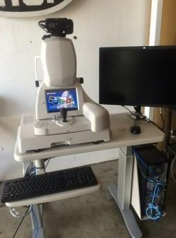
Product Details
In stock 1 Item
Condition Refurbished
| Brand | TOPCON |
| Condition | Refurbished |
| Model | 3D OCT-2000 |
| Warranty | Not specified |
Used Topcon 3D OCT-2000 w/Digital Non-Myd Retinal Camera
The Topcon 3D OCT-2000 System is the first
Spectral Domain OCT system to incorporate a high resolution fundus camera and a userfriendly
color touch screen display in a compact, space saving design.
What makes the Topcon 3D OCT-2000 unique:
» Integrated, high resolution (12.3MP) fundus camera
» FastMap™ software enables dynamic viewing of 2D, 3D and fundus images simultaneously
» Embedded touch-screen for quick and easy navigation
» Historic patient data from Stratus® OCT can be easily imported, analyzed and viewed
» Seamless integration with EyeRoute® Image Management System
3D OCT Image
Topcon’s proprietary FastMap software pioneered the 3D visualization of OCT data, providing another
dimension of clinical information, thereby enhancing the understanding and illustration of complex
pathologies such as vitreous traction, macular edema and retina schesis.
» 3D visualization of OCT data
» Illustrates complex pathologies
» Quickly export images and 3D movies for
presentations
Fundus Image
High resolution, non-mydriatic, color fundus images allow for visualization of conditions which
otherwise would go undetected with OCT technology, such as disc hemorrhages. The 45 degree
field of view and the availability of stereo photos provides the ultimate diagnostic insight.
» High resolution 12.3 MP color fundus camera
» Easily capture and view stereo photos
» PinPoint Registration™ of OCT data in the fundus image
B-scan Image
FastMap software encompasses the latest in noise reduction algorithms and overlapping scanning
technology to create exquisite B-Scan images, which are available almost instantaneously. This
greatly reduces chair time for the patient and enhances offi ce workfl ow.
» Uses latest in noise reduction algorithms
» Enhances office workflow
» Customize capture protocols
Comprehensive Capturing Capture images of the fovea and optic nerve head in one single
scan and high resolution images of the choroid with automatic choroid reference mode.
Video Functions
Use single touch control to review and playback images
and create 2D and 3D videos.
Extended Scanning Depth
Capture high-quality images of high-myopic and hyperopic
patients with a diopter compensation lens and an extended
scanning depth of 2.3 mm.
Compare Function
Allows you to visualize serial exams or view both eyes side by side.
FastMap Software
Topcon’s proprietary FastMap software ensures consistent, high quality images. Using enhanced 3D registration
technology, FastMap reduces artifacts which may be caused by eye movement. This in turn allows for speedy
PinPoint Registration of the OCT data within the fundus image without sacrifi cing workfl ow and preserves image
quality of the vitreo-retinal interface.
In addition, FastMap provides dynamic, simultaneous viewing of the fundus image, and 2D and 3D OCT data,
and automatically detects ILM, RFNL, IS/OS junction, RPE and Bruch’s membrane, which can also be modified.
Thickness Measurement Functions
FastMap software incorporates the latest layer detection algorithms, allowing you to automatically measure total
retinal thickness, RNFL or compare against your legacy Stratus measurements. Manual adjustment of all measurement
grids combined with auto and manual registration of serial exams gives you the highest level of confi dence in
retinal and RFNL thickness measurements.
Mosaic Function
Create panoramic views from the macula to the optic disc.
Specifications
Field Angle 45°
Working Distance 40.7mm
Pupil Diameter ≥ 3.3 mm for Fundus image
Scanning Range 8.2 x 3.0 mm, 6.0 x 6.0 mm, 4.5 x 4.5 mm or 3.0 x 3.0 mm
A Scan Speed 27,000 A Scans/sec.
Scan Depth 2.3mm
Horizontal Resolution 20μm
Longitudinal Resolution 5-6μm
Fundus Observation Near IR
Fundus Camera Nikon D90 12.3 MP Color
Fixation Adjustable internal matrix LCD and external fi xation device
Diopter Scale Range -13D to 12D (in fundus photography)
Light Source Super luminescence diode (SLD)
Wavelength 840nm
Half Bandwidth: 50nm
Output on cornea ≤ 0.65 mW
Automatic OCT Reference Focus Vitreous and Choroid
Scan Patterns 3D, Cross*, Raster*, Line*, Radial*, Circle* (*available with oversampling,
overlapping)
Power Supply Source voltage : AC 100/110/120/220/230/240V 50-60Hz
Power input : 200VA (normal), 400VA (max)
Dimensions 21.5” (W) x 21” (D) x 23.5” – 25” (H)
Weight 70 lbs
Retinal Layers Identifi ed Macula: ILM, IS/OS, RPE, Bruch’s Membrane
Glaucoma: RNFL
Capture PC and Review Station Specifi cations
Platform IBM PC/AT compatible
CPU Pentium 4 or higher
Memory 4GB or higher *2GB or higher for review station
OS Microsoft Windows® XP Professional
Hard Disc 500 GB* or higher *80 GB required for Review Station Software
Display SXGA 1280 x 1024 32-bit color
Graphics Board VRAM 256MB or higher *512MB preferred
Product Details
In stock 1 Item
Condition Refurbished
1
Discuss and agree upon the equipment's condition, configuration, and pricing with the seller before proceeding.
2
Make payment to our secure Escrow account, ensuring funds are held safely until the transaction is complete.
3
Examine the received equipment to confirm its configuration and condition as agreed upon.
4
Once satisfied with the equipment, approve the release of payment to the seller, finalizing the transaction.

USA
Fill Out The Form Below To Open Escrow
Write us an email!
We will call you back soon!
Contact us